
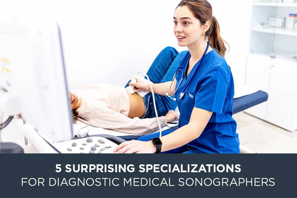
When most people think of medical ultrasound, they immediately visualize an expectant mother viewing an image of her unborn child. While obstetrics remains a well-known and vital application of ultrasound, it represents only a small portion of what ultrasound technology can do. The reality is that ultrasound utilizes high-frequency sound waves to image a vast array of soft tissues, moving organs, and blood vessels throughout the human body. This technical versatility means that the field contains diverse professional paths that many prospective students might not expect.
At MedQuest College, our 24-month Diagnostic Medical Sonography program is available exclusively at our Lexington campus. This program focuses heavily on establishing a solid foundation in Cardiac Sonography and Vascular Sonography. These core disciplines introduce students to the intricate physics of sound and structural anatomy. Once you establish this fundamental knowledge, you can begin to see how broad the field truly is. In this guide, we will explore 5 surprising specializations for diagnostic medical sonographers* that showcase the versatility of this medical imaging path.
1. Musculoskeletal (MSK) Sonography
One of the rapidly growing fields within ultrasound imaging is Musculoskeletal sonography. Historically, physicians relied almost entirely on X-rays or Magnetic Resonance Imaging (MRI) to evaluate joint pain, sports injuries, and soft tissue tears. Today, ultrasound is utilized as a dynamic alternative.
Imaging Tissues in Motion
Unlike static imaging methods, MSK sonography allows the technologist to capture images of the body while it moves. An MSK sonographer might ask a patient to flex a joint, rotate a shoulder, or contract a specific muscle group during the actual scan. This dynamic evaluation helps physicians pinpoint structural issues that only appear when a patient is in motion.
An MSK specialist routinely evaluates:
- Tendons and Ligaments: Checking for tears, strains, or chronic inflammation such as rotator cuff tears in the shoulder or Achilles tendonitis in the ankle.
- Muscles: Locating deep tears, hematomas, or fluid collections within muscle tissue groups.
- Joints: Assessing for the presence of fluid buildup, cysts, or early signs of structural cartilage degradation.
- Foreign Bodies: Assisting physicians in locating small wood, glass, or metal splinters embedded in soft tissue.
This specialized area requires a deep understanding of musculoskeletal anatomy, as the technologist must distinguish between subtle layers of fibers, nerves, and connective tissues.
2. Pediatric Echocardiography
At MedQuest College, our students learn the intricate details of adult Cardiac Sonography, which involves imaging the chambers, walls, and valves of the heart. However, applying these concepts to infants, toddlers, and teenagers requires a specific set of skills known as Pediatric Echocardiography.
Navigating Congenital Heart Anomalies
The primary focus of a pediatric cardiac sonographer is identifying congenital heart defects, which are structural heart issues present at birth. While adult cardiac sonography often focuses on acquired conditions like age-related valve issues or plaque buildup, pediatric sonography requires evaluating hearts that may have formed differently.
A pediatric cardiac sonographer looks for:
- Septal Defects: Identifying openings or holes in the walls that separate the left and right sides of the heart.
- Valve Malformations: Assessing conditions where heart valves are fused, narrowed, or entirely missing.
- Transposition of Great Arteries: Evaluating cases where the primary blood vessels leaving the heart are connected to the wrong chambers.
Because a newborn baby’s heart is roughly the size of a walnut and beats at a rapid rate, this specialization requires physical precision and the ability to operate specialized high-frequency transducers designed for small chest cavities.
3. Ocular Sonography (Ophthalmic Ultrasound)
Perhaps one of the most unexpected specializations for diagnostic medical sonographers involves using ultrasound to look inside the human eye. When structural changes or bleeding prevent an eye care professional from seeing the back of the eye during a standard physical exam, ocular ultrasound becomes an essential diagnostic tool.
Evaluating the Internal Structures of the Eye
Ocular sonographers utilize miniature, highly calibrated probes that are gently placed against a closed eyelid or directly onto the surface of the eye after a numbing agent is applied. This method allows the sonographer to generate real-time cross-sectional images of the eye’s internal components.
This specialty is vital for evaluating:
- Retinal Detachment: Determining if the delicate light-sensitive tissue at the back of the eye has separated from its underlying support layers.
- Vitreous Hemorrhage: Detecting blood collections within the clear gel that fills the center of the eye cavity.
- Intraocular Tumors: Assisting oncologists in measuring the exact dimensions and characteristics of masses inside the globe of the eye.
- Foreign Objects: Locating microscopic debris that may have penetrated the eye during industrial accidents or trauma.
4. Fetal Echocardiography
While general obstetrical ultrasound looks at the overall growth of a fetus, Fetal Echocardiography is a highly detailed sub-specialty dedicated exclusively to analyzing the fetal heart. This scan is typically requested when a routine pregnancy ultrasound detects a potential irregularity or when a family history increases the likelihood of a congenital heart issue.
Detailed Screening Before Birth
A fetal echocardiographer performs a meticulous evaluation of the heart structures while the fetus is still inside the womb, often during the second trimester of pregnancy. The sonographer must track the flow of blood through tiny vessels that are fractions of a millimeter wide.
Key areas of focus during a fetal echocardiogram include:
- Four-Chamber View: Confirming that all four chambers of the heart are proportional and forming correctly.
- Outflow Tracts: Ensuring that blood is moving in the correct direction into the aorta and pulmonary arteries.
- Fetal Heart Rhythms: Documenting and analyzing irregular heartbeats or arrhythmias that may require medical intervention immediately following delivery.
This path requires a strong grasp of both fetal circulatory paths and complex wave physics, as the sonographer must navigate maternal tissue and fetal positioning to get clear structural data.
5. Neurosonology (Neonatal and Adult Brain Imaging)
Using sound waves to look through bone may sound counterintuitive, but Neurosonology is a well-established specialty that allows sonographers to evaluate the neurological structures and blood flow patterns within the brain.
Neonatal Neurosonology
In newborn infants, the bones of the skull have not yet completely fused, leaving soft areas known as fontanelles. Neonatal sonographers can use these natural acoustic windows to place an ultrasound probe directly on an infant’s head, generating clear images of the internal brain tissue without exposing the vulnerable patient to ionizing radiation. This is commonly performed in neonatal intensive care units to check for intraventricular bleeding or structural developmental changes.
Adult Transcranial Doppler (TCD)
In adult patients, the skull is fully fused, but sonographers can still evaluate the brain using Transcranial Doppler (TCD) ultrasound. By placing the ultrasound probe against specific thin areas of the skull, such as the temple or the base of the neck, the sonographer can track the velocity of blood moving through the primary cerebral arteries. This technique is useful for monitoring patients who have experienced a stroke, assessing vasospasms following a brain aneurysm, or screening individuals with sickle cell disease for specific neurological risks.
The Role of Educational Foundations in Choosing a Path
When evaluating diagnostic medical sonographer specializations, it is important to remember that all of these pathways rely on the same core principles of physics, anatomy, and patient care. No matter which specific branch of ultrasound catches your interest, you must begin with a structured educational program.
Building Your Foundation at MedQuest College
MedQuest College provides a 24-month Associate of Applied Science Degree path at our Lexington campus. Our program focuses on Cardiac and Vascular sonography, which are technically demanding areas of medical imaging.
Our structured program includes:
- On-Campus Lectures and Labs: Spend time understanding ultrasound physics, hemodynamics (the mechanics of blood flow), and cross-sectional anatomy.
- Hands-on Training: Practice using current ultrasound machines in our clinical labs under the direct guidance of experienced instructors.
- Clinical Externships: Complete real-world rotations in local hospitals and outpatient centers to apply your skills in active medical settings.
By gaining competency in cardiac and vascular imaging, you build the critical thinking skills and technical dexterity needed to adapt to alternative specializations later in your educational and professional journey.
Frequently Asked Questions (FAQs)
Q: Can I specialize in multiple types of ultrasound at the same time?
A: Yes. Many sonographers hold multiple credentials through credentialing bodies such as the American Registry for Diagnostic Medical Sonography (ARDMS) or Cardiovascular Credentialing International (CCI). Holding credentials in multiple areas, such as vascular technology and cardiac sonography, allows you to work in a broader range of clinical environments.
Q: How long does it take to learn these different specializations?
A: The educational journey begins with an accredited associate degree program, which takes 24 months to complete at our Lexington campus. Once you graduate and earn your initial credentials, you can pursue secondary certifications through additional coursework, clinical training hours, or specialized examination pathways.
Q: Does MedQuest College offer institutional accreditation for its sonography program?
A: Yes. MedQuest College is institutionally accredited by the Accrediting Bureau of Health Education Schools (ABHES). Our Diagnostic Medical Sonography program also holds programmatic accreditation through the Commission on Accreditation of Allied Health Education Programs (CAAHEP), ensuring our curriculum meets rigorous national standards.
Q: Is there a significant amount of math and physics involved in ultrasound training?
A: Yes. Ultrasound equipment relies on complex wave principles, the Doppler effect, and fluid dynamics. A portion of your 24-month program will be dedicated to understanding ultrasound physics so you can adjust equipment settings to capture clear diagnostic images safely.
Learning About the Field
The applications of ultrasound go well beyond standard expectations. Specialized ultrasound practices allow professionals to image tissues in motion, trace complex cardiovascular abnormalities in infants, and evaluate vascular flow within the brain. Understanding the 5 surprising specializations for diagnostic medical sonographers underscores the versatility of this technology in modern patient care.
If you are ready to take the first step toward building a foundation in diagnostic imaging, exploring the curriculum at MedQuest College is a practical starting point. Our dedicated faculty and hands-on laboratory structures are designed to assist you as you navigate your educational milestones.
Are you ready to tour our Lexington scanning labs? Visit our Diagnostic Medical Sonography program page to request additional information, review specific admissions requirements, or connect with our financial aid advisors today.
*Institutional Note on Specializations and Credentialing: While the medical imaging field includes diverse clinical applications, not all specialized scanning techniques exist as standalone professional credentials. Some competencies, such as Transcranial Doppler (TCD), are integrated as required graduation skills within foundational pathways like Vascular Technology. Other historical pathways, including Neurosonology, are now sub-specialties evaluated under broader, general registry examinations. Specialized pathways like Ocular Sonography are regulated by independent ophthalmic boards with separate, specific prerequisites. MedQuest College provides the foundational Associate of Applied Science curriculum in Cardiac and Vascular Sonography, which serves as the academic starting point for these broader clinical applications.